

Project – Time Reversal Beamforming Imaging for Breast Cancer Detection
- Project Description: Microwave radiation is well known as a diagnostic imaging method for many medical applications, for example, early state breast cancer detection. Microwave detection of breast tumors is a non-ionising, potentially low cost, in vivo modality that relies on the dielectric contrast between healthy and malignant breast tissues. The scattering environment in a breast often appears to be inhomogeneous due to changing dielectric properties of the beast tissues. Such an environment imposes challenges to existing imaging methods. Time reversal (TR) is an adaptive waveform transmission scheme that utilizes the rich scattering medium to best match to the target response. This project uses time reversal to increase the detectability of malignant tumors with higher resolution. This project involves algorithm development and programming of imaging methods; experimental collecting of electromagnetic field data; and 2-dimensional or 3-dimensional finite difference time domain simulation modeling.
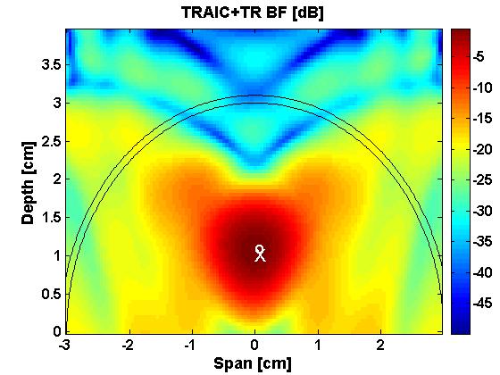

- Fig.1. Left figure shows the TR image of a breast tumor; Right figure shows the direct subtraction image. The cross indicates the exact location of the target, and the circle indicates the peak of the image. The left figure has a high contrast indicating a better resolution.
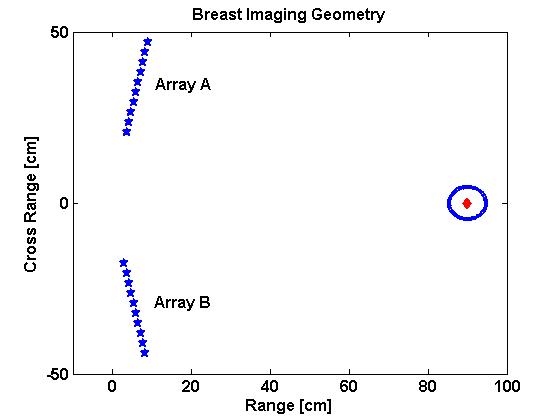
- We carry our electromangetic feasibility study of the time reversal beamforming imager. The experimental setup is illustrated in Fig.7. The left panel shows the imaging geometry. The horizontal axis denotes the range; the vertical axis denotes the cross range. The diamond denotes the target location; the circle represents the skin layer of a breast phantom. Two antenna arrays, A and B, are placed with an angle of 11.3 radians relative to the vertical axis. Both the arrays and the breast phantom are stationary. Thus, we obtain measured data from fixed look angles by the two arrays.

- Fig.-2: Experimental feasibility of time reversal beamforming imager. We use a highly simplified breast phantom that consists of a PVC pipe, a wood dowel or a glass rod, and Styrofoam (polystyrene). The PVC pipe represents the breast skin; the wood dowel and the glass rod represent the tumor targets; and the Styrofoam fills the PVC, representing the breast tissues.
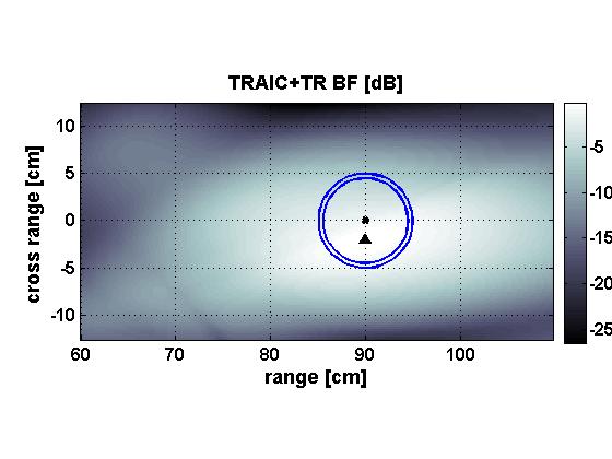

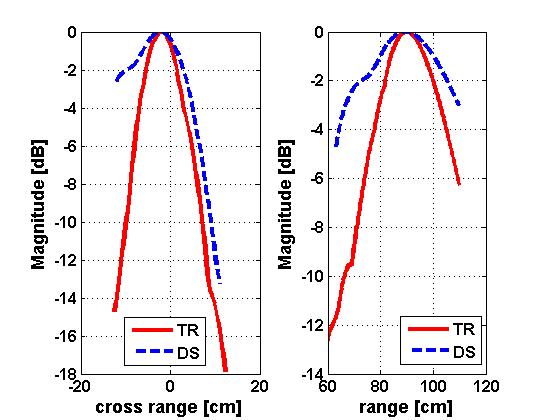
- Fig.-3. Wood tumor imaging results. Left — TR method; Right — Direct subtraction method. The two images are scaled to the same dynamic range for comparison purposes. The sharper and higher contrast image has more details and thus a finer resolution. In both figures, the dot denotes the tumor location; the triangle denotes the estimated tumor location. The results show that the location estimation error for time reversal imaging is smaller than the direct subtraction method. The following figures shows the projection of the two images in cross range (left) and the projection in the range direction (right). Examining the -3 dB main lobe width reveals that time reversal yields a narrower beamwidth, i.e., a finer resolution, than the direct subtraction method.
Selected Publications:
- Y. Jin, J. M. F. Moura, Y. Jiang, M. Wahl, and Q. He, “Breast cancer detection by time reversal imaging”, 5th IEEE International Symposium on Biomedical Imaging: From Nano to Macro, ISBI’08, Paris, Friance, May 14-17, 2008 [ Full-Text]
- Y. Jin, Y. Jiang, and J.M.F. Moura, “Time reversal beamforming for microwave breast cancer detection”, IEEE International Conferences on Image Processing, September 16-19, San Antonio, Texas, 2007 [ Full-Text]